8 February 2026
Why a microscope changes everything in root-canal treatment
Most failed root-canals fail for one reason: a missed canal. Working under a microscope at 16× magnification turns that statistic on its head.
Dt. Müberra BayraktarFounder · Oral Implantology Specialist

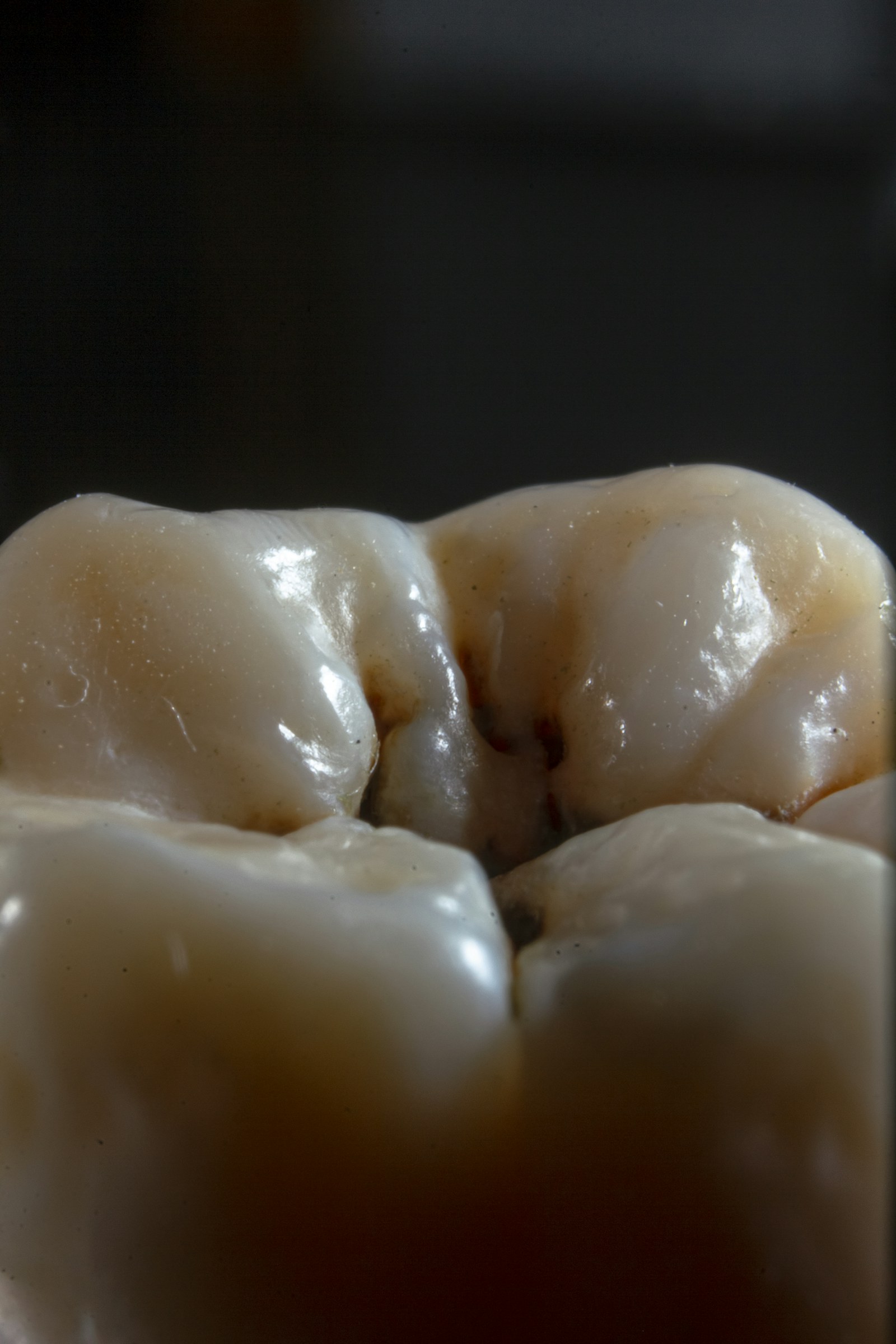
Conventional endodontics is done either with the naked eye or with simple magnifying loupes (2.5–4×). At that resolution, narrow accessory canals — especially the MB2 canal in upper molars — are easily missed. A missed canal continues to harbour bacteria, and the treatment fails months or years later.
Under a surgical microscope at 12–16× with coaxial fibre-optic illumination, the picture changes. We can see calcifications, hairline fractures, and the dark dot of an unexpected canal mouth that would otherwise be invisible.
That single difference is why our endodontic success rate on first-attempt molar treatments is above 95%, against an international average closer to 80% for naked-eye work.
If you've been told your tooth is 'not savable', it is worth a second opinion under the microscope before extraction.
Continue reading